
The Role of Medical Imaging in Diagnosis and Care

TL;DR:
- Medical imaging is essential for diagnosing, monitoring, and guiding treatment of medical conditions by creating internal body visuals. It is a core part of healthcare that supports clinical decisions, treatment planning, and ongoing management across various techniques like X-ray, CT, MRI, and ultrasound. Proper use of imaging involves stewardship to balance benefits and risks, including radiation exposure and incidental findings, while technological advances like AI and molecular imaging are shaping a proactive, personalized future.
Medical imaging is the process of creating visual representations of the inside of the body to diagnose, monitor, and guide treatment of medical conditions. The role of medical imaging in modern healthcare is foundational. An estimated 340 million contrast-enhanced X-ray, CT, and MRI procedures are performed annually worldwide. That number reflects how central imaging has become to everyday clinical decisions, from detecting a broken bone to identifying early-stage cancer. Understanding what these technologies do, and when they are truly needed, helps you become a more informed participant in your own care.
What is the role of medical imaging in healthcare?
Medical imaging is classified as part of the information-gathering phase of diagnosis. Its primary purpose is to narrow diagnostic possibilities and reduce clinical uncertainty without requiring surgery or invasive procedures. When your doctor orders an imaging test, the goal is to see what cannot be felt or heard during a physical exam.
The importance of medical imaging extends well beyond the initial diagnosis. Imaging also guides treatment decisions. A surgeon planning to remove a tumor needs to know its exact size and location before making a single incision. An oncologist monitoring a patient’s response to chemotherapy relies on repeat scans to measure whether the tumor is shrinking. Imaging is not a one-time event. It is a continuous tool throughout a patient’s care pathway.
Imaging also plays a direct role in managing chronic conditions. Patients with heart disease, lung disease, or diabetes often receive periodic scans to track organ function and catch complications early. At Gardenstatemedicalgroup, diagnostic imaging services are integrated into primary care and specialty programs precisely because ongoing monitoring matters as much as the first diagnosis.
What are the main medical imaging techniques?
Modality selection follows a clear hierarchy based on the clinical question, the body part involved, and the patient’s safety profile. Each technique works differently and serves a distinct purpose.
X-ray is the first-line tool for quick imaging of bones and lungs. It uses low doses of ionizing radiation to produce flat, two-dimensional images. A chest X-ray can detect pneumonia, rib fractures, or an enlarged heart in minutes. X-rays are fast, widely available, and cost-effective for straightforward clinical questions.
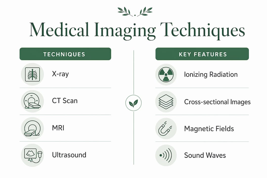
CT (computed tomography) scans take X-ray technology further by producing detailed cross-sectional images of the body. CT is the preferred tool for urgent imaging, such as evaluating trauma injuries or detecting internal bleeding. The trade-off is a higher radiation dose compared to a standard X-ray.
MRI (magnetic resonance imaging) uses magnetic fields and radio waves rather than radiation. It produces the clearest images of soft tissues, including the brain, spinal cord, muscles, and ligaments. MRI exams typically last between 30 and 60 minutes, and safety screening for metal implants is required before every scan. MRI is the gold standard when soft-tissue detail is the clinical priority.
Ultrasound uses sound waves to create real-time images of organs and blood flow. It involves no radiation, making it the preferred choice for imaging during pregnancy and for evaluating abdominal organs. Ultrasound is also used to guide needle biopsies and drain fluid collections in real time.
The table below summarizes the key differences across these four core techniques.
| Technique | How it works | Best used for | Radiation involved |
|---|---|---|---|
| X-ray | Ionizing radiation | Bones, lungs, quick screening | Yes (low dose) |
| CT scan | Multiple X-ray angles | Trauma, internal organs, emergencies | Yes (moderate dose) |
| MRI | Magnetic fields and radio waves | Brain, spine, soft tissues | No |
| Ultrasound | Sound waves | Pregnancy, abdomen, real-time guidance | No |
How does imaging support diagnosis, treatment, and monitoring?
Medical imaging in diagnosis works by giving clinicians objective visual evidence to confirm or rule out a condition. A patient presenting with chest pain could have a pulmonary embolism, a rib fracture, or a cardiac event. Imaging separates these possibilities quickly and accurately. Without it, clinicians rely solely on symptoms and lab results, which are often insufficient on their own.
Treatment planning depends heavily on imaging findings. Radiation oncologists use CT and MRI scans to map the exact boundaries of a tumor before designing a radiation treatment field. Orthopedic surgeons review X-rays and MRIs before deciding whether a fracture requires surgery or a cast. The images do not just confirm a diagnosis. They shape the entire treatment strategy.
Monitoring disease progression is equally important. A patient recovering from a stroke receives follow-up MRI scans to assess brain recovery. A patient with a lung nodule receives periodic CT scans to watch for growth. Imaging guides treatment at every stage, not just at the point of initial diagnosis.
Clinical practice is also shifting toward personalized imaging protocols tailored to individual patient needs rather than standardized, one-size-fits-all approaches. This means the type of scan, the timing, and the imaging parameters are increasingly matched to the specific patient and condition. The result is more accurate information with less unnecessary exposure.
Pro Tip: Before any imaging test, ask your doctor two questions: what specific finding will this scan change, and what happens if we wait? These questions help you and your provider decide whether the scan is truly necessary right now.
What are the risks and limits of medical imaging?
Medical imaging is not without downsides. Low-value imaging can cause emotional harm, unnecessary costs, radiation exposure, and further invasive testing triggered by incidental findings. An incidental finding is something spotted on a scan that was not the reason for the scan, and it often requires additional testing to rule out a serious problem, even when the original finding turns out to be harmless.
Overutilization is a real and documented problem. Clinicians increasingly order imaging to manage diagnostic uncertainty and reduce liability, not always because the scan will change the clinical plan. This drives unnecessary testing and exposes patients to risks that outweigh the benefits in those specific cases.
Imaging stewardship is the practice of balancing the benefits and risks of each scan to avoid unnecessary testing. Patients and providers share responsibility for this. You can ask whether a scan is truly needed. Your provider can apply evidence-based criteria before ordering one. Together, these conversations reduce low-value imaging without compromising care quality.
MRI safety deserves specific attention. Strong magnetic fields make MRI unsafe for patients with certain metal implants, pacemakers, or cochlear devices. MRI screening for implants is mandatory before every scan. For patients with claustrophobia or anxiety, wide-bore MRI machines and sedation options are available to make the experience manageable.
Preparation also matters more than most patients realize. Scan preparation varies by modality and can include fasting, drinking contrast agents, or stopping certain medications. Arriving unprepared can delay or invalidate the test entirely. Always confirm your preparation instructions with your provider’s office before the appointment.
Pro Tip: If you have any metal implants, a pacemaker, or a history of surgeries involving devices, tell your imaging team before your MRI appointment. This information is critical for your safety and must be reviewed before the scan begins.
What does the future of medical imaging look like?
The future of medical imaging moves well beyond producing clearer pictures. Molecular imaging complements anatomical imaging by visualizing biological processes at the cellular level. This enables earlier disease detection and more precise characterization of conditions like cancer, before structural changes are even visible on a standard scan.
Artificial intelligence is reshaping how radiologists interpret images. AI systems can flag abnormalities, measure tumor volumes, and compare current scans to prior studies in seconds. This supports faster, more consistent diagnoses and reduces the chance of a subtle finding being missed. The integration of AI with radiomics, which extracts large amounts of data from imaging pixels, is opening the door to predictive modeling that goes far beyond what the human eye can detect alone.
The benefits of medical imaging are also expanding through individualized patient approaches that match imaging protocols to each patient’s specific biology and risk profile. A 40-year-old with a family history of colon cancer may follow a different imaging schedule than a 40-year-old without that history. Personalized protocols reduce unnecessary scans while catching the conditions most likely to affect that specific patient.
Early risk detection through advanced imaging is shifting medicine from reactive to proactive. Conditions caught earlier are almost always easier and less costly to treat. Imaging is increasingly the tool that makes that early detection possible.
Key Takeaways
Medical imaging is the most direct, non-invasive method clinicians have for seeing inside the body, and its value depends entirely on using the right technique for the right clinical question at the right time.
| Point | Details |
|---|---|
| Imaging serves multiple roles | Imaging supports diagnosis, treatment planning, and ongoing disease monitoring throughout patient care. |
| Technique selection matters | X-ray, CT, MRI, and ultrasound each serve distinct clinical purposes based on body area and safety profile. |
| Overuse carries real risks | Low-value imaging causes radiation exposure, emotional harm, unnecessary costs, and incidental findings. |
| Stewardship is a shared responsibility | Patients and providers should discuss whether each scan will change the clinical plan before ordering it. |
| The field is advancing rapidly | Molecular imaging and AI are shifting imaging from a diagnostic tool to a clinical decision driver. |
Why imaging stewardship changed how I think about scans
When I first started paying close attention to how imaging is ordered and used, I expected the story to be simple: more imaging equals better care. The reality is more nuanced, and honestly more interesting. The shift toward imaging stewardship is not about doing less. It is about doing the right thing at the right time for the right patient.
What strikes me most is how often patients arrive expecting a scan, and how rarely they ask what the scan will actually change. That one question, “what will this result change about my treatment?” is the most powerful tool a patient has in any imaging conversation. Providers who welcome that question are the ones practicing medicine at its best.
The advances in molecular imaging and AI are genuinely exciting. But the technology only delivers value when it is applied thoughtfully. A faster, sharper image of the wrong thing at the wrong time is still the wrong decision. The future of imaging is not just better machines. It is better judgment about when and how to use them.
— Krunal
Imaging services at Gardenstatemedicalgroup
Gardenstatemedicalgroup offers radiology and diagnostic testing services designed to support accurate diagnosis and ongoing care management for patients in North Bergen and Secaucus, New Jersey.
The practice integrates imaging with primary care, cardiopulmonary services, and chronic disease programs so that your scan results connect directly to your broader care plan. Whether you need a chest X-ray, an MRI, or a specialized diagnostic procedure, the team at Gardenstatemedicalgroup works to match the right imaging tool to your specific clinical needs. Contact Gardenstatemedicalgroup to schedule an appointment or ask about which diagnostic procedures are appropriate for your situation.
FAQ
What is the role of medical imaging in diagnosis?
Medical imaging is classified as part of the information-gathering phase of diagnosis, designed to narrow diagnostic possibilities and reduce clinical uncertainty. It gives clinicians objective visual evidence to confirm or rule out conditions without invasive procedures.
What are the most common medical imaging techniques?
The four primary techniques are X-ray, CT scan, MRI, and ultrasound. Each works differently and is selected based on the body area, clinical question, and the patient’s safety profile.
Is medical imaging safe?
Most imaging techniques are safe when used appropriately. MRI involves no radiation but requires screening for metal implants. CT and X-ray involve ionizing radiation, which is why clinicians weigh the benefit against the exposure risk before ordering these scans.
What is imaging stewardship?
Imaging stewardship is the practice of ordering scans only when the results will meaningfully change the clinical plan. It reduces unnecessary radiation exposure, costs, and incidental findings that can lead to further invasive testing.
How should I prepare for a medical imaging appointment?
Preparation varies by scan type and may include fasting, stopping certain medications, or drinking a contrast agent. Always confirm your specific instructions with your provider’s office before the appointment to avoid delays or rescheduling.